Global Institute Of Stem Cell Therapy And Research
US: +1-800-914-7836

What's new

Latest news

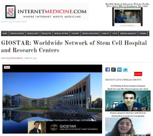
GIOSTAR: Worldwide Network of Stem Cell Hospital and Research Centers Coverage on InternetMedicin.com

GIOSTAR: Worldwide Network of Stem Cell Hospital and Research Centers

(ED NOTE: There is no other area of Healthcare that sorely need both healthcare worker education, and patient education, than the field of Stem Cells; there seems to be an unlimited source of promise, and an area that needs continuing research to mine this tremendous promise.  This entire post is taken from the website of this organization.  The San Diego office is a Research Center)

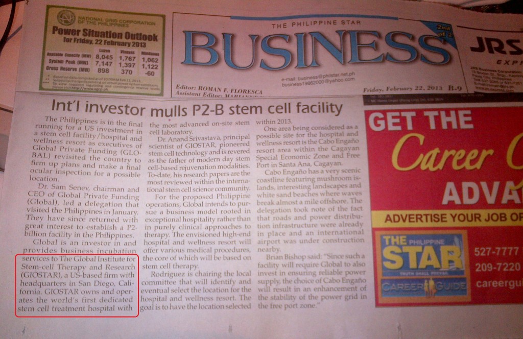
Global Institute of Stem Cell Therapy and Research (GIOSTAR), headquartered in San Diego, California (U.S.A.), was formed with the vision to provide stem cell based therapy to aid those suffering from degenerative or genetic diseases around the world.

We are the leaders in developing cutting edge stem cell based technology, supported by leading scientists with the pioneering publications in the area of stem cell biology. Our primary focus is to discover and develop a cure for human diseases with innovative and unique stem cell based therapies and products. Regenerative medicine promises treatment for those diseases previously regarded as incurable. GIOSTAR’s ultimate aim is to maintain its leadership in the field of stem cell science and to continue developing affordable stem cell based therapy for those in need.

In July 2011, GIOSTAR inaugurated the world’s first dedicated stem cell treatment hospital at Ahmedabad, Gujarat, India. This is a 125-bed state of the art facility with the most advanced on-site stem cell laboratory.

GIOSTAR has an ability to treat several devastating Immunological diseases and Blood related diseases. These include Diabetes Type I, Lupus, Multiple Sclerosis, Crohns, Vasculitis, Scleroderma, Myasthenia Gravis, Sickle cell anemia, Leukemia, Lymphoma, Thalassemia and developing the therapies for Alzheimer’s, Autism, Anti-Aging Treatments, Parkinson’s, , Cancer, Heart and Retinal Degeneration, Amyyotrophic Lateral Sclerosis, Neuropathy, Osteoarthritis, Paralysis, Strokes, Spinal Cord Injuries, Skin Burns and Spinal Muscular Atrophy (SMA).

GIOSTAR is in process of opening dedicated stem cell treatment hospitals in Asia, Middle East, Africa, North and South America and Europe. The proposed facilities will be developed with the international support from public and private sectors grants and investments to provide stem cell based therapy for those suffering from degenerative or genetic diseases.

GIOSTAR uses autologous and allogeneic adult stem cells transplant to treat patients, following the highest medical standards.

GIOSTAR is leading the most advanced research to use embryonic stem (ES) cells and Induced Pluripotent Stem (IPS) cells to develop new therapies for clinical use.

GIOSTAR is a coalition of esteemed and illustrious scientific minds in the field of genetic and stem cell science. For over 15 years GIOSTAR team of scientists and clinicians have been involved in the development and utilization of stem cell based clinical protocols related to stem cell transplants.

The GIOSTAR team includes international leaders in the field of embryonic stem (ES) cells and Induced Pluripotent Stem (IPS) cells research and technologies. GIOSTAR team is the first in demonstrating the significance of ES cell use for the development of therapies for several degenerative diseases related to tissue and organs. The publications of these therapies have been thoroughly investigated and documented by many noted journals of medicine. GIOSTAR cutting-edge Stem Cell science will be used to treat qualified patients on needed basis.

GIOSTAR is using three kinds of stem cells : adult stem cell, human embryonic stem cell and Induced Pluripotent cells. The adult stem cell technology is well developed and being used regularly in clinical practice. Our clinicians are licensed to treat the patients using autologous (their own) and allogeneic (from others) adult stem cells for certain diseases. We are the leading private organization, which has state of art excellence in extraction of stem cell from a human body and use it for therapy. Our clinicians are treating the patients regularly using adult stem cells since 2000. GIOSTAR is leading the most advanced research to use embryonic stem (ES) cells and Induced Pluripotent Stem (IPS) cells to develop new therapies for clinical use.

Basal Expression of Pluripotency-Associated Genes Can Contribute to Stemness Property and Differentiation Potential

Basal Expression of Pluripotency-Associated Genes Can Contribute to Stemness Property and Differentiation Potential


Nidheesh Dadheech,1 Abhay Srivastava,1 Muskaan Belani,1 Sharad Gupta,2 Rajarshi Pal,3 Ramesh R. Bhonde,3Anand S. Srivastava,4 and Sarita Guptacorresponding author1


Associated Data

Supplementary Materials

Abstract

Pluripotency and stemness is believed to be associated with high Oct-3/4Nanog, and Sox-2 (ONS) expression. Similar to embryonic stem cells (ESCs), high ONS expression eventually became the measure of pluripotency in any cell. The threshold expression of ONS genes that underscores pluripotency, stemness, and differentiation potential is still unclear. Therefore, we raised a question as to whether pluripotency and stemness is a function of basal ONS gene expression. To prove this, we carried out a comparative study between basal ONS expressing NIH3T3 cells with pluripotent mouse bone marrow mesenchymal stem cells (mBMSC) and mouse ESC. Our studies on cellular, molecular, and immunological biomarkers between NIH3T3 and mBMSC demonstrated stemness property of undifferentiated NIH3T3 cells that was similar to mBMSC and somewhat close to ESC as well. In vivo teratoma formation with all three germ layer derivatives strengthen the fact that these cells in spite of basal ONS gene expression can differentiate into cells of multiple lineages without any genetic modification. Conclusively, our novel findings suggested that the phenomenon of pluripotency which imparts ability for multilineage cell differentiation is not necessarily a function of high ONS gene expression.

Introduction

Ability to isolate and establish pluripotent or multipotent stem/precursor population is a major objective for the biotechnological industry and clinical translation of regenerative medicine. A major impediment for using stem cells in a clinical setup is poor availability of cells, especially those obtained with noninvasive procedures without raising much ethical issues. These limitations greatly restrict the usage of stem cells in clinics, disabling treatments of many degenerative diseases. This lacuna can be filled if any tissue-specific cells can be verifiably demonstrated to possess pluripotent or multipotent capacity. This may elevate the hope to find a well-suited stem cell-like cell line that can serve as an autologous, noncontroversial, and renewable source for cell therapy without ethical and immunological concerns, which are usually associated with embryonic stem cells (ESCs).

Numerous gene and protein expression criteria have been set for recognizing a cell as pluripotent. Microarray analyses have demonstrated a set of various transcripts that are associated with stemness as in the case of ESCs [1]. Notably, it has been demonstrated by Yamanaka and colleague that the combinations of four major transcription factors, Oct-3/4NanogSox-2, and Klf-4, is indispensable for the cells to maintain pluripotency as in the case of ESCs [2,3]. However, there are some other reports which indicate that expressions of not all the four genes are essential to maintain the stemness. According to NIH and ISSCR guidelines, teratoma formation is one of the major criteria for classifying a cell to be pluripotent [4]. Apart from this, the presence of alkaline phosphatase (ALP) is another reliable property shown for pluripotent cells such as ESCs [1,5].

In principle, a cell that is able to differentiate into cell types of all three germinal layers is considered pluripotent. On the other hand, a multipotent cell can give rise to cells originating from the same germ layer [6]. A classic example of a stem cell with pluripotent and multipotent potential is mesenchymal stem cells (MSC). MSCs can differentiate into various cell types. Depending on isolation procedure and tissue source, both pluripotent and multipotent type of stem cells can be isolated [7]. Mesenchymal cells display fibroblastic cell morphology and express vimentin protein as a characteristic marker along with high Oct-3/4Nanog, and Sox-2 (ONS) expression [8]. Bone marrow MSC (BMSC) isolated from crude bone marrow are reported to possess pluripotent gene expression and also show tri-lineage differentiation [912]. Therefore, it appears that BMSC can serve as a model that is used for screening various cell types for their degree of stemness. Hence, cells which portray specific features that are in accordance with the parameters mentioned earlier can be designated as pluripotent cells.

In a very recent report, Wang et al. exemplify that ONS genes are the core regulators of pluripotency. This group showed that Oct4 regulates and interacts with the BMP4 pathway to specify four developmental fates. High levels of Oct4 enable self-renewal in the absence of BMP4; Nanog represses embryonic ectoderm differentiation but has little effect on other lineages, whereas Sox-2 is redundant and represses mesendoderm differentiation. Thus, instead of being panrepressors of differentiation, each factor controls specific cell fates [13]. Numerous cell types isolated from tissues and organs such as heart, kidney, liver, lungs, brain, pancreas, spleen, muscles, adipose tissue, dental pulp, placenta, and amnion are now being tested on the parameters mentioned earlier with an aim of generating patient-specific pluripotent stem cell lines for treating various incurable degenerative diseases.

With numerous published reports, it becomes a general belief that pluripotency, stemness, and differentiation potential into trigerminal lineages are associated with high levels of ONS genes, but this does not explain the fact that many low ONS expressing cells can also demonstrate multilineage differentiation potential, high ALP staining, and a teratoma-like structure formation and, hence, generate valid questions. (1) What should be the threshold of ONS gene expression for any cell to be pluripotent? (2) Is pluripotency really a function of high ONS gene expression or can basal level expressing cells also show lower but sustained pluripotency? Attempts are made in the present study to answer these questions scientifically using NIH3T3 as a model and are compared with high ONS expressing cells.

NIH3T3 cells, isolated from Swiss albino mice [14], are not known as stem cells due to the reduced expression of pluripotency-associated genes despite being embryonic in nature. These cells have been extensively used as a model system for various molecular, cellular, and toxicological studies [1518] and are considered differentiated dermal fibroblast cells. Many groups used NIH3T3 as control cells that fail to differentiate [1921]. However, their differentiation into adipocytes, myocytes, and neural cell types has been reported either after transforming genetically or if differentiated under defined media conditions [1921]. Piestun et al. reported that NIH3T3 cells after being transfected with Nanog show induced pluripotency and expressed markers that are specific to various differentiating cell types [22]. In 2006, Yamanaka and colleagues also compared pluripotent ESCs and untransformed NIH3T3 by a microarray analysis and concluded that although these cells did not express pluripotency genes, especially the four crucial ones, that is, Oct3/4NanogSox-2, and Klf4; they expressed c-Myc [3]. However, this group neither examined nor commented on the multipotent nature of these cells. Later, in 2008 and 2009, Deng and colleagues published two reports demonstrating differentiation of untransformed NIH3T3 cells into oesteocytes and neuronal cell types in bone and spinal cord injury mouse models [1,23,24]. Two other groups also reported that NIH3T3 can give rise to neural cells and oesteoblasts, respectively [21,25]. Recently, our group also reported pancreatic islet cell differentiation of untransformed NIH3T3 with appropriate induction [26].

To answer these questions and to understand whether pluripotency can also be associated with basal ONS gene expression, we carried out a comparative study of basal ONS expressing NIH3T3 cells with pluripotent mouse BMSC (mBMSC) and mouse ESC (mESC). We carefully examined NIH3T3 cells for pluripotent and multipotent stem cell specific markers and compared them with those of mBMSC and mESC with regard to morphological features, expression profile of stemness related genes, ability to undergo multilineage differentiation, and in vivo teratoma formation.

Materials and Methods

Animals

Male Balb/c mice, 3–4 weeks old, weighing around 25–30 g, were used for transplantation and bone marrow cell isolation experiments. All animal experiments were performed in accordance with our institutional ‘‘Ethical Committee for Animal Experiments’’ and CPCSEA guidelines and regulations.

Chemicals and cell culture media

All chemicals, media, and antibodies used in this study were purchased from Sigma Aldrich. Molecular biology reagents and cDNA and PCR kits were procured from Fermentas Inc. All plasticwares were purchased from Nunc, Inc.

Isolation of marrow-derived MSCs

Bone marrow-derived mononuclear cells were isolated from 4 week-old Balb/c mice according to Zhu et al. [10,27]. Mice were sacrificed after proper anaesthesia. Briefly, tibia and femur bones were dissected under sterile conditions. The metaphyseal ends of the bones were cut, and the marrow plugs were flushed out by passing low glucose Dulbecco’s modified Eagle’s medium (L-DMEM) (Sigma) through a needle inserted into one end of the bone. Bone marrow cells from mice were collected; big tissue debris were removed with the help of a cell strainer and were then seeded in low-glucose RPMI-1640 medium supplemented with 10% fetal bovine serum (FBS) for 2 days.

Purification and propagation of MSCs

MSCs were purified as described earlier [10,27]. Briefly, the cultured cells were seeded at a density of 10,000 cells/mL in complete medium (high glucose DMEM supplemented with 10% FBS (Sigma), penicillin (100 IU/mL), streptomycin (100 IU/mL; MP Biological, Inc.) in polystyrene T25 culture flasks and incubated in a humidified 5% CO2 incubator at 37°C for 48 h. Loosely attached hematopoietic cells were discarded on trypsinizing adherent cells for 5 min. Cells that remained adhered were trypsinized for another 7 min [27] and then cultured in complete medium for 10 days. The cell media was replaced with fresh one every third day. The cells were trypsinized using Trypsin-EDTA (Sigma-Aldrich, USA) and passaged in T75 flasks until the second generation became 70%–90% confluent. Cultured mBMSC were then characterized and used for a comparative study with NIH3T3 after ensuring their pluripotency.

Cell culture and maintenance

NIH3T3 cells were obtained from three different sources (Cell Repository, National Centre for Cell Science, Pune, India, ATCC procured cells from Zydus Pharmaceuticals, Ahmedabad, India and a generous gift from Dr. Girish M. Shah’s lab, Laval University. Quebec, Canada) and were maintained in high-glucose DMEM supplemented with 10% FBS. Cells were split into a 1:1 ratio in new tissue culture flasks on being about 90% confluent. Culture media was changed after every 2 days.

Cell count and growth curve

Fully confluent mBMSC and NIH3T3 cells were trypsinized with 0.1% Trypsin EDTA solution and counted under an inverted phase-contrast microscope (Nikon TE2000). One hundred thousand cells were seeded into each well of 24-well plates for growth curve studies. Cells were eventually trypsinized and counted at different time points (0, 24, 48, 72, 96, and 120 h). Cell counts were then plotted versus time to establish the growth curve of cells. Growth curve of NIH3T3 cells was compared with that of mBMSC. Doubling time of both the cells were also determined using the algorithm ln (Nt−N0) ln (t), where Nt and N0 were number of cells at final time point and at initial seeding point, respectively, and t was time period in hours for which cell counts were recorded.

MTT [3-(4,5-dimethylthiazol-2-yl)-2,5 diphenyltetrazolium bromide] assay

Cell proliferation rate and effect of cytotoxic agent [dimethyl sulphoxide (DMSO)] were determined by MTT assay (Roche). NIH3T3 cells were seeded into 96-well plates at a density of 50,000 cells per well in complete medium. MTT reagent was added in culture in each well after 24 h of incubation with different concentrations of DMSO. The optical density values were analyzed 4 h after the MTT reaction using Multiskan PC (Thermo Lab), and cell survival curves were then plotted against time in culture.

H&E staining

Cells were trypsinized and fixed in 4% paraformaldehyde (PFA) in phosphate-buffered saline (PBS) solution. A histological examination was carried out by standard histological techniques. Fixed cells were stained with H&E stain, and observations were recorded under a light microscope.

Transfection and GFP labeling

Before carrying out an in vivo teratoma assay, we transfected NIH3T3 cells with GFP harboring plasmid p-eGFP-N1 (Clontech), so that the cells could be differentiated from those of the host. One hundred thousand cells were transfected with 1 μg p-eGFP-N1 plasmid DNA using lipofectamine 2000 (Invitrogen) as a transfecting agent in a 1:3 volume ratio in a 3.5 cm2 dish, according to the manufacturer’s protocol. Following the transfection, stably GFP expressing clones were selected by growing them in media containing G418 at 300 μg/mL concentration for the first 2 days and thereafter in 900 μg/mL G418 for the next 7 days. Clones were purified from isolated colonies using clonal discs and scaled up for subcutaneous transplants with agarose plugs in Balb/c mice.

Teratoma formation

For each graft, ∼0.2 million GFP expressing NIH3T3 cells, washed and resuspended in 300 μL DMEM complete medium, were injected subcutaneously into Balb/c mice (maintained in MSU in-house animal house facility) in 1.5% melted agarose [2830]. Visible tumors, after 2 weeks of transplantation, were dissected and fixed overnight with 4% PFA solution. The tissues were then paraffin embedded, sectioned, stained with H&E, and, the sections were examined for the presence of cells that were representatives of all three germ layers produced by NIH3T3-eGFP cells [31].

ALP staining

ALP staining of spheroids of both NIH3T3 and mBMSC origin was carried out with BCIP/NBT stain. Cell clusters were mechanically isolated, and cells were dissociated with trypsin EDTA digestion for 5 min at 37°C. After washing with 10% FBS and then with PBS, the cells were fixed with 2% PFA solution for 5 min and stained with NBT/BCIP [1]. 3T3L-1 cells served as controls.

Immunocytochemistry

An immunocytochemical analysis was performed for various stem/progenitor markers in both cell types as previously described [32,33]. Cells were seeded on glass coverslips in high-glucose DMEM complete medium and allowed to adhere for 3–4 h. After attachment, cells were fixed with 4% PFA solution for 10 min at room temperature. After permeabilizing them with 0.1% Triton X-100 in PBS for 5 min, the cells were incubated in blocking buffer (1% bovine serum albumin and 4% FBS in PBS) for 1 h at room temperature. Primary antibodies were added in blocking solution, and coverslips were incubated overnight at 4°C. A list of the antibodies used and their dilutions are given in Table 1. Cells were then washed thrice with washing buffer (1:10 dilution of blocking buffer in PBS) and incubated with secondary antibodies conjugated to FITC and TRITC flurophores (Sigma Aldrich) in the dark for 30 min at room temperature. For negative controls, cells were incubated with normal IgG. Cells were mounted with Vectashield mounting medium containing 4′6′-diamidino-2-phenylindole dihydrochloride (DAPI) (Vector Laboratories, Inc.). Fluorescence imaging was carried out by using a Nikon TES2000 microscope. All images were analyzed with the help of NIH element software supplied by Nikon Japan.

Read More : Click Here

GIOSTAR Executives Invited to Congressional Luncheon

Global Institute of Stem Cell Therapy and Research (GIOSTAR) Chairman Dr. Anand Srivastava and CEO Mr. Deven Patel were among the invited guests at the San Diego Regional Chamber of Commence hosted Congressional Luncheon on April 19, 2013. This is one of San Diego’s best opportunities to hear directly from the San Diego regional congressional delegation and to exchange the ideas with leadership.

Among the congressional attendees, Congresswoman Susan Davis, Congressman Duncan D. Hunter, Congressman Darrell Issa, Congressman Scott Peters, Congressman Juan Vargas were present at the luncheon.

GIOSTAR Chairman Dr. Anand Srivastava had a long talk with Congressman Scott Peters regarding visa problem faced by the Professionals in the scientific communities in San Diego area.  These highly qualified scientists immigrating from India, China and other parts of the world are not getting their conclusive immigrant status after working for seven to ten years at the local institutions and contributing great deal in the advancement of scientific research.

Congressional Luncheon 1 Congressional Luncheon 2 Congressional Luncheon 3 Congressional Luncheon 4

Congresswoman Susan Davis and GIOSTAR CEO Mr. Deven Patel discussed small business grants in the biotechnology field and finding a ways to boost local biotech business.  “San Diego being very strong incubator for biotech research, it can not affords to loose its ground due to lack of funding,” said Mr. Patel.

Today GIOSTAR scientists study all three types of stem cells: tissue-derived stem cells, embryonic stem (ES) cells and induced pluripotent stem (iPS) cells, as well as progenitor cells. Molecular biology, genetic engineering and protein chemistry are core technologies for conducting this research. We consider all of these cell types as viable options both as cell-based therapies and as tools and technologies for drug discovery and development.

Dr. Anand Srivastava said he had very fruitful conversation with Congressman Juan Vargas for developing entire Baha California region for biotech and research industry.

Congressman Vergas was very open to the idea for developing entire region and make San Diego the epicenter for all developments.

GIOSTAR Executives Invited to Congressional Luncheon

Global Institute of Stem Cell Therapy and Research (GIOSTAR) Chairman Dr. Anand Srivastava and CEO Mr. Deven Patel were among the invited guests at the San Diego Regional Chamber of Commence hosted Congressional Luncheon on April 19, 2013. This is one of San Diego’s best opportunities to hear directly from the San Diego regional congressional delegation and to exchange the ideas with leadership.

Among the congressional attendees, Congresswoman Susan Davis, Congressman Duncan D. Hunter, Congressman Darrell Issa, Congressman Scott Peters, Congressman Juan Vargas were present at the luncheon.

GIOSTAR Chairman Dr. Anand Srivastava had a long talk with Congressman Scott Peters regarding visa problem faced by the Professionals in the scientific communities in San Diego area.  These highly qualified scientists immigrating from India, China and other parts of the world are not getting their conclusive immigrant status after working for seven to ten years at the local institutions and contributing great deal in the advancement of scientific research.

Congresswoman Susan Davis and GIOSTAR CEO Mr. Deven Patel discussed small business grants in the biotechnology field and finding a ways to boost local biotech business.  “San Diego being very strong incubator for biotech research, it can not affords to loose its ground due to lack of funding,” said Mr. Patel.

Today GIOSTAR scientists study all three types of stem cells: tissue-derived stem cells, embryonic stem (ES) cells and induced pluripotent stem (iPS) cells, as well as progenitor cells. Molecular biology, genetic engineering and protein chemistry are core technologies for conducting this research. We consider all of these cell types as viable options both as cell-based therapies and as tools and technologies for drug discovery and development.

Dr. Anand Srivastava said he had very fruitful conversation with Congressman Juan Vargas for developing entire Baha California region for biotech and research industry.

Congressman Vergas was very open to the idea for developing entire region and make San Diego the epicenter for all developments.

GIOSTAR Executives Invited to Congressional Luncheon Giostar2 Giostar3 Giostar4

Therapeutic potential of mesenchymal stem cells in regenerative medicine

Therapeutic potential of mesenchymal stem cells in regenerative medicine

Devang M. Patel, 1 Jainy Shah, 1 and Anand S. Srivastava 2 ,*


Abstract

Mesenchymal stem cells (MSCs) are stromal cells that have the ability to self-renew and also exhibit multilineage differentiation into both mesenchymal and nonmesenchymal lineages. The intrinsic properties of these cells make them an attractive candidate for clinical applications. MSCs are of keen interest because they can be isolated from a small aspirate of bone marrow or adipose tissues and can be easily expanded in vitro. Moreover, their ability to modulate immune responses makes them an even more attractive candidate for regenerative medicine as allogeneic transplant of these cells is feasible without a substantial risk of immune rejection. MSCs secrete various immunomodulatory molecules which provide a regenerative microenvironment for a variety of injured tissues or organ to limit the damage and to increase self-regulated tissue regeneration. Autologous/allogeneic MSCs delivered via the bloodstream augment the titers of MSCs that are drawn to sites of tissue injury and can accelerate the tissue repair process. MSCs are currently being tested for their potential use in cell and gene therapy for a number of human debilitating diseases and genetic disorders. This paper summarizes the current clinical and nonclinical data for the use of MSCs in tissue repair and potential therapeutic role in various diseases.

1. Introduction

Stem cells are immature tissue precursor cells which are able to self-renew and differentiate into multiple cell lineages [12]. Mesenchymal stem cells (MSCs), also known as multipotent mesenchymal stromal cells, are self-renewing cells which can be found in almost all postnatal organs and tissues [34]. MSCs have received wider attention because they can be easily isolated from a small aspirate of bone marrow or adipose tissue and can be expanded to clinical scales in in vitro condition. Other than these MSCs offer several other advantages like long-term storage without major loss of potency and no adverse reactions to allogeneic MSCs transplant [5].

In 1976 Friedenstein et al. firstly described a method for MSCs (referred as “stromal cells”) isolation from whole bone marrow aspirates based on differential adhesion properties. They suggested that these cells are adherent, clonogenic, nonphagocytic, and fibroblastic in nature, with the ability to give rise to colony forming units-fibroblastic (CFU-F) [6]. In late 1980s Owen and Friedenstein reported heterogeneity of the bone marrow stromal cells for the first time [78]. Bone marrow stromal cells were further characterized and named mesenchymal stem cell to describe the subtype of marrow stromal cells involved in the process of mesengenesis [910]. Shortly after these discoveries researchers started to explore the therapeutic application of MSCs [11], since then no adverse effect of MSC transplantation has been reported. In this paper we tried to compile recent advances in the MSCs research and its medical implications.

2. Immunophenotype of MSC

The identification of MSCs with the use of specific markers remains elusive. There is no single surface marker, but rather a panel of surface markers which define Human MSCs (hMSCs), derived from fresh tissues or cryopreserved samples. As per the international society for cellular therapy guidelines, MSCs must express CD105 (SH2), CD73 (SH3/4), and CD90 and must be negative for surface markers CD34, CD45, CD14, CD79α or CD19, and HLA-DR [9]. hMSCs are also negative for several other antigens like CD4, CD8, CD11a, CD14, CD15, CD16, CD25, CD31, CD33, CD49b, CD49d, CD49f, CD50, CD62E, CD62L, CD62P, CD80, CD86, CD106 (vascular cell adhesion molecule [VCAM]-1), CD117, cadherin V, and glycophorin A. On the other hand, hMSCs are positive for CD10, CD13, CD29 (b1-integrin), CD44, CD49e (a5-integrin), CD54 (intercellular adhesion molecule [ICAM]-1), CD58, CD71, CD146, CD166 (activated leukocyte cell adhesion molecule [ALCAM]), CD271, vimentin, cytokeratin (CK) 8, CK-18, nestin, and von Willebrand factor [51213]. Tissue specific expression of surface marker is well noted such as only adipose tissue-derived MSCs express high levels of CD34 [14] and bone-marrow-derived MSCs, but not placenta derived MSCs, express CD271 [15]. Detailed phenotypic expression of surface markers is reviewed elsewhere [16].

3. Differentiation Potential of MSC

Other than surface markers MSCs must have ability to adhere to plastic and differentiate into osteoblasts, adipocytes, and chondroblasts under in vitro condition [9]. Differentiation is regulated by genetic events, involving transcription factors. Differentiation to a particular phenotype pathway can be controlled by some regulatory genes which can induce progenitor cells’ differentiation to a specific lineage. Besides growth factors and induction chemicals, a microenvironment built with biomaterial scaffolds can also provide MSCs with appropriate proliferation and differentiation conditions [17]. Even though MSCs can differentiate into a number of tissues in vitro, the resulting cell population does not mimic the targeted tissues entirely in their biochemical and biomechanical properties [18].

3.1. Mesoderm Differentiation

Theoretically, mesodermal differentiation is easily attainable for MSCs because they are from same embryonic origin. In the literature also mesoderm (osteogenic, adipogenic, and chondrogenic) differentiation is relatively well studied. A mixture of Dexamethasone (Dex), β-glycerophosphate (β-GP), and ascorbic acid phosphate (aP) has been widely used for induction in osteogenic differentiation [1819]. Osteogenic differentiation of MSCs is a complex process that is tightly controlled by numerous signaling pathways and transcription factors [20]. Runt-related transcription factor 2 (Runx2) and Caveolin-1 are considered a key regulator of osteogenic differentiation which is precisely regulated by numerous activators and repressors [1921]. Bone morphogenetic proteins (BMPs), especially BMP-2, BMP-6, and BMP-9, have been shown to enhance osteogenic differentiation of MSCs [18]. Smads, p38 and Extracellular signal-Regulated Kinase-1/2 (ERK1/2) are involved in BMP9-induced osteogenic differentiation [22]. At very low concentration BMP-2, vascular endothelial growth factor (VEGF) and basic fibroblast growth factor (bFGF) synergistically promote the osteogenic differentiation of rat bone marrow-derived mesenchymal stem cells. Other than core binding factor alpha-1/osteoblast-specific factor-2 (cbfa1/osf2) [23], Wnt signaling has also been implicated in osteogenic differentiation of MSCs [24]. Recently a study by Alm et al. showed that transient 100 nM dexamethasone treatment reduces inter- and intraindividual variations in osteoblastic differentiation of bone marrow-derived human MSCs [25]. An alternative approach would be to use a scaffold or matrix engineered to provide cues for differentiation. Silicate-substituted calcium phosphate (Si-CaP) supported attachment and proliferation of MSCs was proved to be osteogenesis [26]. In adipogenesis differentiation, Dex and isobutyl-methylxanthine (IBMX) and indomethacin (IM) have been used for induction and have been observed by staining the lipid droplets in cells by Oil Red O solution. Peroxisome proliferator-activated receptors-γ2 (PPAR-γ2), CCAAT/enhancer binding protein (C/EBP), and retinoic C receptor have been implicated in adipogenesis [17]. Phosphatidylinositol 3-kinase (PI3 K) activated by Epac leads to the activation of protein kinase B (PKB)/cAMP response element-binding protein (CREB) signaling and the upregulation of PPARγexpression, which in turn activate the transcription of adipogenic genes, whereas osteogenesis is driven by Rho/focal adhesion kinase (FAK)/mitogen-activated protein kinase kinase (MEK)/ERK/Runx2 signaling, which can be inhibited by Epac via PI3 K [27].

In chondrogenesis differentiation, transforming growth factor (TGF)-β1 and TGF-β2 are reported to be involved [28]. Differentiation of MSCs into cartilage is characterized by upregulation of cartilage specific genes, collagen type II, IX, aggrecan, and biosynthesis of collagen and proteoglycans. The emerging results suggested the possible roles of Wnt/β-catenin in determining differentiation commitment of mesenchymal cells between osteogenesis and chondrogenesis [19]. A recent report suggested that miR-449a regulates the chondrogenesis of human MSCs through direct targeting of Lymphoid Enhancer-Binding Factor-1 [29]. Elevated β-catenin signaling induces Runx2, resulting in osteoblast differentiation, whereas reduced β-catenin signaling has the opposite effect on gene expression, inducing chondrogenesis [30]. Fibroblasic Growth factor-2 (FGF-2) can enhance the kinetics of MSC chondrogenesis, leading to early differentiation, possibly by a priming mechanism [31].

3.2. Ectoderm Differentiation

In vitro neuronal differentiation of MSCs can be induced by DMSO, butylated hydroxyanisole (BHA), β-mercaptoethanol, KCL, forskolin, and hydrocortisone [17]. Moreover, Notch-1 and protein kinase A (PKA) pathways are found to be involved in neuronal differentiation [32]. In presence of other stimulatory, downregulation of caveolin-1 promotes the neuronal differentiation of MSCs by modulating the Notch signaling pathway [33].

3.3. Endoderm Differentiation

In liver differentiation, hepatocyte growth factor and oncostatin M were used for induction to obtain cuboid cells which expressed appropriate markers (α-fetoprotein, glucose 6-phosphatase, tyrosine aminotransferase, and CK-18) and albumin production in vitro [34]. Recent studies identified methods to develop pancreatic islet β-cell differentiation from adult stem cells with desirable results. The resulting cells showed specific morphology, high insulin-1 mRNA content, and synthesis of insulin and nestin [3536]. Murine adipose tissue-derived mesenchymal stem cells can also differentiate to endoderm islet cells (expressing Sox17, Foxa2, GATA-4, and CK-19) with high efficiency then to pancreatic endoderm (Pancreatic and duodenal homeobox 1[Pdx-1], Ngn2, Neurogenic differentiation [NeuroD], paired box-4 [PAX4], and Glut-2), and finally to pancreatic hormone-expressing (insulin, glucagon, and somatostatin) cells [37].

4. Migration and Homing

The physical niche and migration signals of MSCs provide invaluable information about their role and interactions within the tissue. Bone-marrow-derived MSCs received more attention from researchers in hopes of revealing clues about their therapeutic activity. During in vivo condition, it is difficult to locate MSCs’ niche. Moreover due to the lack of any specific MSCs marker and difficulties in probing marrow cavities, it is very difficult to track dynamic movement of MSC. Most researchers use genetic markers such as Y-chromosome, when male cells are introduced into females or fluorescent protein reporter genes but these methods do not resolve the dynamics of cellular and temporal responses and are not quantitative [5]. Noninvasive in vivo imaging accomplished by using bioluminescence imaging (BLI) can be a possible solution. The main advantage of BLI is that even at very low levels of signal, as few as 100 cells can be detected in vivo [3839]. Significant advances have been made in this field but still MSCs migration to tissue niche is illusive.

MSCs migration to injured tissues has been reported in radiation-induced multiorgan failure, ischemic brain injury, myocardial infarction, and acute renal failure [40], but the mechanisms that regulate the MSCs migration to the injured tissues are still unknown. Human MSCs express different combinations of the chemokine receptors CCR1, CCR4, CCR7, CCR9, CCR10, CXCR1, CXCR3, CXCR4, CXCR5, and CX3CR1 [41]. The chemokine(s) that control MSCs trafficking are still unknown; while to date, 39 chemokines have been identified with different functions controlling the traffic of hematopoietic cells, in particular leukocytes [41]. Among these chemokines, stromal cell derived factor-1 (SDF-1) is relatively well studied for MSCs migration.

SDF-1-induced cell migration is mediated by its receptor, CXCR4, which is broadly expressed in cells of the immune system and in the central nervous system (CNS). The role of SDF-1 as an important mediator of stromal progenitor migration to injured tissue has been reported in vivo using a rat model of myocardial infarction [4243]. Hiasa et al., 2004 reported that the overexpression of human SDF-1 in the ischemic muscle induced the mobilization of endothelial progenitor cells and improved myocardial healing. Studies also demonstrated that after myocardial infarction the levels of SDF-1 are increased in infarcted tissue and this increase correlates with the number of MSCs that home into the heart [4243]. On the other hand, study by Ip et al., 2007 suggested that MSCs use integrin β1 and not CXCR4 for their myocardial migration [44]. Moreover, in regenerating skeletal tissues, the MSCs homing may be improved with growth factor delivery, as combined MSCs and erythropoietin infusion gave better results in limb ischemia treatment [45]. Bioactive lipid lysophosphatidic acid (LPA) plays a principal role in the migration of human lung resident MSCs through a signaling pathway involving LPA1-induced beta-catenin activation [46]. Anti-inflammatory environment is more accommodating to the therapeutic hMSCs than a proinflammatory environment [47].

Crossing of the endothelial barrier is another critical step for the tissue migration of circulating cells. Similarly to leukocytes, MSCs adhesion to the endothelial cells represents a critical step and a restricted set of molecules such as selectin-P, integrin β1, and VCAM-1 and seems to play critical roles in this interaction [48]. The in vivo homing potential of MSCs circulating in the bloodstream to the sites of injury/inflammation can be regulated by adhesion of MSCs to endothelium, achieved by pretreatment of endothelial cells with some proapoptotic agents, angiogenic and inflammatory cytokines, and growth factors, such as interleukin (IL)-8, neurotrophin-3, TGF-β, IL-1β, TNF-α, platelet-derived growth factor, EGF, and SDF-1 [12]. Further studies into understanding the molecular mechanism behind migration and homing will provide an impetus to the use of MSCs for therapeutic purpose.

5. Mechanism of Action/Mode of Action

The mechanism by which MSCs exert their antiproliferative effect have still to be fully elucidated, although several mechanisms and molecules have been proposed that are likely to act in concert and/or in alternate fashion depending on the environment conditions to which MSCs are exposed. Several studies have shown that MSCs are capable of replacing damaged tissues in vivo [4950]. Multiple tissue engineering approaches have also been reported where undifferentiated or predifferentiated MSCs were delivered with or without help of biomaterial [4950]. MSCs have shown promise in replacing various tissues including cartilage, bone, tendon, vasculature, liver kidney, and nerve [51]. However, it remains unclear that how many originally delivered MSCs retain residency in the wounded tissue and maintain the appropriate terminally differentiated phenotype because large amount of transplanted population become apoptotic within the initial phase, or migrate to lungs and liver. Study on stroke and cardiac injury by Li et al. and Askari et al., respectively, suggested that transient MSCs presence appears to be sufficient to elicit a therapeutic effect [5253]. Taking together these findings suggests that resident MSCs also work to suppress both transient and perpetual immune surveillance systems and create an ideal healing environment by secreting factors and altering the local microenvironment [51].

Since 2002 in vitro T-lymphocyte activation and proliferation assays have been used in several studies which resulted in understanding the immunomodulatory effect of MSCs from human, murine, and baboon [5456]. These studies demonstrated that MSCs were capable of suppressing both lymphocyte proliferation and activation in response to allogeneic antigens. Moreover, MSCs can induce development of CD8+regulatory T (Treg) cells than can in turn successfully suppress allogeneic lymphocyte responses [56] and prohibit differentiation of monocytes and CD34+ progenitors into antigen presenting dendritic cells [57]. T cells stimulated in presence of MSCs get arrested in the G1 phase as a result of cyclin D2 downregulation [58]. MSCs are also capable of inhibiting the proliferation of IL-2 or IL-15 stimulated NK cells [5960]. MSCs have also been shown to alter B-cell proliferation, activation, IgG secretion, differentiation, antibody production, and chemotactic behaviors [51]. Treatment with in vitro expanded allogeneic MSCs successfully resolved severe grade IV acute graft-versus host disease (GvHD) supported in vivo immunomodulatory properties of MSCs [61]. Furthermore, MSCs reduce expression of major histocompatibility complex class II (MHCII), CD40, and CD86 on Dendritic cell (DC) following maturation induction [51]. Interestingly, allogeneic MSCs which were differentiated towards a chondrogenic phenotype continued to suppress antigen specific T-cell proliferation in rheumatoid arthritis [62] and genetically engineered MSCs escaped immune rejection and induced ectopic bone formation in vivo [56]. However several other reports suggested that the immunomodulatory effects of MSCs are not universal and unconditional and that the MSCs phenotype is transient and context dependent [63].

Cytokine secretion is one of the major therapeutic characteristics of MSCs [64]. MSCs secretion is not limited to factors like TGF-β, IL-10, IL-6, cyclooxygenase-1 (COX-1), and COX-2 which are responsible for prostaglandin E2 (PEG2) secretion. MSCs partly inhibited DC differentiation through IL-6 secretion and reduced tissue inflammation by IL-10, TGF-β1, and IL-6 secretion [5765]. TGF-β1 secretion by MSCs suppresses T-lymphocyte proliferation and activation, initiated by IL-1β secretion from CD14+monocytes [66]. In fact one study suggested that only the supernatants obtained from cocultures of stromal cells and activated T cells displayed an immunosuppressive effect when added to secondary cultures of proliferating T cells [5867]. Taking together MSCs mediated immunosuppression is not exclusively the result of a direct inhibitory effect but involves the recruitment of other regulatory effects. Details about immune-modulation of immune response are reviewed elsewhere [6869].

Read More : Click Here

Make An Inquiry

We are here to help you

WordPress Video Lightbox