Global Institute Of Stem Cell Therapy And Research
US: +1-800-914-7836

Category

Stem Cell News

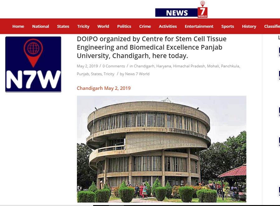
DOIPO organized by Centre for Stem Cell Tissue Engineering and Biomedical Excellence Panjab University, Chandigarh

DOIPO organized by Centre for Stem Cell Tissue Engineering and Biomedical Excellence Panjab University, Chandigarh


A special lecture on Stem Cell Sciences by Dr. Sahiba Singh, Stem Cell Physician, Global Institute of Stem cell therapy and Research (GIOSTAR), USA

Chandigarh May 2, 2019

A special lecture on Stem Cell Sciences by Dr. Sahiba Singh, Stem Cell Physician, Global Institute of Stem cell therapy and Research (GIOSTAR), USA along with Dr. Joydeep Das Gupta, Director of India Project Operation organized by Centre for Stem Cell Tissue Engineering and Biomedical Excellence Punjab University, Chandigarh, here today.  They explained the use of Stem Cell therapy for the treatment of degenerative disease, such as Parkinson disease, Alzheimer’s disease, and Diabetes etc. The have also explained the various successful clinical case studies by the use of  Stem Cell therapy.  Lecture on Stem Cell Therapy was attended by M.Sc. students Research Scholars & faculty Members.  “

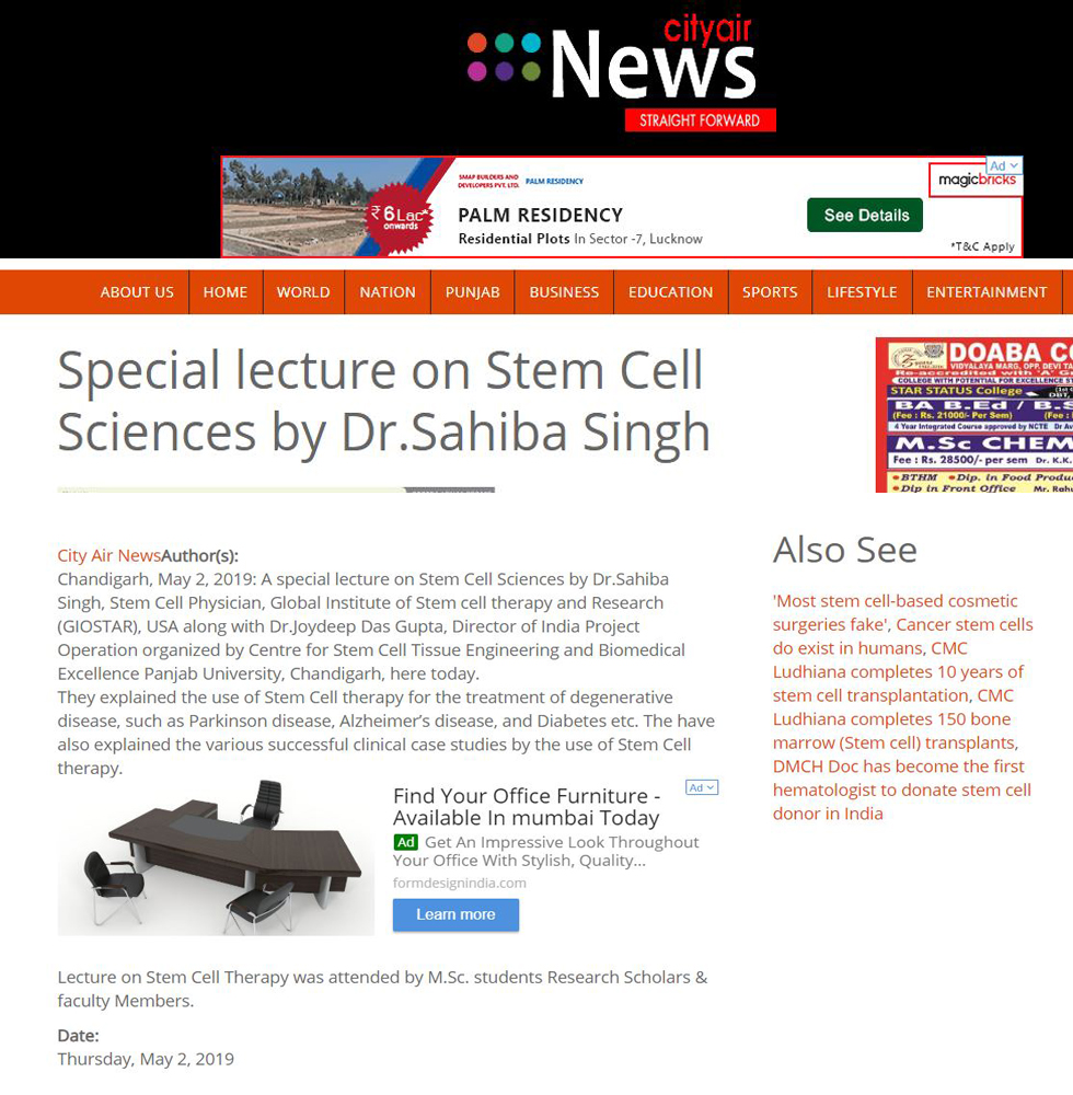
Special lecture on Stem Cell Sciences by Dr. Sahiba Singh

Special lecture on Stem Cell Sciences by Dr. Sahiba Singh – City Air News


Special lecture on Stem Cell Sciences by Dr.Sahiba Singh

Chandigarh, May 2, 2019:

A special lecture on Stem Cell Sciences by Dr. Sahiba Singh, Stem Cell Physician, Global Institute of Stem cell therapy and Research (GIOSTAR), USA along with Dr. Joydeep Das Gupta, Director of India Project Operation organized by Centre for Stem Cell Tissue Engineering and Biomedical Excellence Panjab University, Chandigarh, here today.

They explained the use of Stem Cell therapy for the treatment of degenerative disease, such as Parkinson disease, Alzheimer’s disease, and Diabetes etc. The have also explained the various successful clinical case studies by the use of Stem Cell therapy.

Lecture on Stem Cell Therapy was attended by M.Sc. students Research Scholars & faculty Members.

Mesenchymal Stem Cell Conditioned Media Ameliorate Psoriasis Vulgaris: A Case Study

Mesenchymal Stem Cell Conditioned Media Ameliorate Psoriasis Vulgaris: A Case Study


Seetharaman R, Mahmood A, Kshatriya P, Patel D, Srivastava A. Case Reports in Dermatological Medicine. May 2019; Article ID 8309103. https://doi.org/ 10.1155/ 2019/ 8309103.

Case Reports in Dermatological Medicine

Volume 2019, Article ID 8309103, 5 pages
https://doi.org/10.1155/2019/8309103
Case Report

Mesenchymal Stem Cell Conditioned Media Ameliorate Psoriasis Vulgaris: A Case Study

1GIOSTAR Research Inc. Pvt. Ltd., Ahmedabad, Gujrat, India
2Global Institute of Stem Cell Therapy and Research, 4660 La Jolla Village Drive, San Diego, CA 92122, USA

Correspondence should be addressed to Anand Srivastavaanand@giostar.com

Received 6 April 2019; Accepted 21 April 2019; Published 2 May 2019

Academic Editor: Jacek Cezary Szepietowski

Copyright © 2019 Rajasekar Seetharaman et al. This is an open access article distributed under the Creative Commons Attribution License, which permits unrestricted use, distribution, and reproduction in any medium, provided the original work is properly cited.

Abstract

Psoriasis, an autoimmune disease, affects a vast number of peoples around the world. In this report, we discuss our findings about a scalp psoriasis suffering patient with a Psoriasis Scalp Severity Index (PSSI) score of 28, who was treated with Mesenchymal stem cell conditioned media (MSC-CM). Remarkably, complete regression was recorded within a treatment period of one month only (PSSI score of 0). A number of bioactive factors like cytokines and growth factors secreted by MSCs in the conditioned medium are very likely to be the principle molecules which play a vital role in skin regeneration. Treatment using MSC-CM appears to be an effective tool for tackling the psoriatic problem and, thus, may offer a new avenue of therapy which could be considered as an alternative approach to overcome the limitations of the cell-based therapy.

1. Introduction

Psoriasis is a chronic disease thought to be of autoimmune origin which is characterized by patches on the skin and nails. It has been considered as a serious skin related problem affecting approximately 100 million individuals worldwide. About 2% of the world population and 0.44-2.8% of the Indian population were affected by psoriasis in 2016-2017 [12]. Plaque, guttate, inverse, pustular, and erythrodermic are the five major types of psoriasis. Plaque psoriasis, also known as psoriasis vulgaris, is the most common form of the disease (about 90% of the cases) [3] which typically presents with red patches with white scales on top. Psoriasis vulgaris which commonly affects the areas includes scalp, knees, elbows, hands, nails, and feet [4].

Psoriasis, an autoimmune-inflammatory disease probably predisposed due to genetic makeup, is mediated by T-helper cells. Polymorphism, referred to as differences in DNA sequences of a gene, can be incurred by various external agents like chemicals, viruses, or radiation. Polymorphisms in genes of Th2 cytokine/regulatory T-cell (interleukin-10/IL10), Th1/Th17 cytokine (IL-12B and IL-23R), and tumour necrosis factor alpha (TNFAIP3; TNIP1) confer which increased other risks like cardiovascular diseases amongst psoriasis patients [57]. Single nucleotide alteration caused polymorphism in Th1 proinflammatory cytokine gene IL-2 [–330 (G/T)] which has been shown to be associated with greater disease severity in the Indian population [1]. On the other hand, another gene polymorphism occurring in Th-2 cytokine/regulatory T-cell (IL-4) has been shown to be protective against psoriasis [5]. Upregulation in the levels of inflammatory cytokines leads to psoriasis which also can be associated with an increased risk of psoriatic arthritis, lymphomas, cardiovascular risk, Crohn’s disease, and depression [3]. There is no permanent cure for psoriasis, though steroid creams, vitamin D3 cream, ultraviolet light, and immune system suppressing medications (methotrexate) have been in wide use to help control the symptoms with some success [89].

Mesenchymal stem cells (MSCs) are multipotent adult stem cells which have an excellent capacity to proliferate for an extended period of time while maintaining the undifferentiated cell status. The resulting daughter cells can differentiate into various types of cells of host tissues and thus help repair wear and tear incurred [10]. MSCs have a potential to serve as a powerful tool in cell-based therapy due to their tissue regenerative and host immune modulatory capabilities. The functions exhibited by MSCs have attracted a number of scientists and clinicians to investigate the mechanisms involved in their curative and tissue regeneration functions. A very few articles have reported the effectiveness of stromal vascular fraction (SVF)/MSC therapy in curing psoriasis by regulating the immune systems. Lee et al. [11] reported that human umbilical cord blood-derived mesenchymal stem cells (hUCB-MSCs) ameliorate psoriasis-like skin inflammation in mice and have regulatory effects on immune cells including CD4+ T cells and dendritic cells. The first case study on intravenous infusion of SVF into psoriasis patient demonstrated a significant decrease in symptoms with a noticeable difference in skin appearance, psoriasis area, and severity index (PASI) score reduction (from 50.4 to 0.3) [12]. Chen et al. [13] reported that umbilical cord-derived MSC (UC-MSC) infusion effectively reduced psoriasis in human subjects. It was believed that MSCs’ migration into the skin lesions and their immunomodulatory, autoimmune inhibitory, and paracrine effects were the principal factors behind the ameliorative effects. Other recent preclinical studies have shown that stem cell-derived conditioned media (CM) exhibit effective healing of psoriasis-like wounds and thus CM is an alternative for several cell-based therapies [1415]. The paracrine factors including growth factors, chemokines, and cytokines secreted from stem cells play a major role in wound healing [16] and these molecules are present in CM or spent medium harvested from cultured cells [17]. In short, CM can serve as a novel treatment approach in regenerative medicine which has been shown to have a successful outcome in preclinical studies. However, a very few reports are available on the clinical application of CM for treatment of any disease. Based on the principles and importance of CM, the present study was aimed at investigating the effect of MSC-CM on a patient suffering from psoriasis. This study is believed to be the first clinical report on the use of MSC-CM to treat psoriasis.

2. Case Report

2.1. Patient

A 38-year-old male patient, who was suffering from psoriasis vulgaris for 2 years, paid a visit to our centre. Preliminary examination of the patient showed that numerous erythematous plaques with numerous silvery scales present all over the scalp including the area behind the ears. The severity of the disease was assessed to be 28 on Psoriasis Scalp Severity Index (PSSI), calculated by the standard method which combines the severity (erythema, induration, and desquamation) and percentage of affected area.

2.2. Preparation of MSC-Conditioned Media

Adipose tissue was collected from a healthy volunteer by lipoaspiration by a plastic surgeon under the aseptic conditions in the O.T. About 100 ml of fat was aspirated out from the waist area and collected in a sterile container. The fat tissue contacting stem cells was processed in a biosafety laminar airflow chamber. MSCs were isolated from adipose tissue by standard enzymatic digestion method with 0.1% collagenase type I. Following the centrifugation, the resulting pellet was cultured in DMEM medium (Invitrogen, Paisley UK) supplemented with 10% foetal bovine serum (FBS) and 1% penicillin/streptomycin, at 37°C in humidified atmosphere containing 5% CO2. The media were changed after every 3 days. About 5×10⁶ MSCs of passage 2 were seeded in each T175 culture flask (n=10) containing 30 ml of DMEM medium supplemented with 10% FBS. MSCs were confirmed with spindle shaped morphology and free from any contamination (Figure 1) using a phase-contrast microscope. When cells attained 90% confluence at passage 2, the culture media were replaced with serum-free DMEM. After 72 h of incubation, resulting MSC-CM was collected, centrifuged at 2000 rpm for 5 min to remove the cell debris, filtered through 0.22-μm filter, and then concentrated (10 times) by ultrafiltration using centrifugal filtering units with a cut-off value of 3 kDa (Amicon Ultra-15; Millipore, MA), according to the manufacturer’s instructions. The concentrated MSC-CM was aliquoted and stored at -20°C until use. MSC-CM was topically applied on the afflicted areas once a day over a period of one month. Clinical parameters like severity, changes, and clearance of psoriatic plaques were monitored at regular intervals.

Figure 1: Phase-contrast microscopic image showing spindle shaped MSCs (× 100).
2.3. MSC-CM Ameliorates Psoriasis Vulgaris

Numerous psoriatic erythematous plaques with adhering silvery scales over the different regions of the scalp were observed before the treatment regimen started. In general, the number of the scales declined significantly within 2 weeks of topical application of MSC-CM. Interestingly, clearance of silvery scales and severity of psoriatic plaques were completely abolished within one month of the treatment (Figure 2). The PSSI score reduced to 0 from 28 and regression of the disease continued for 6 months of follow-up. The patient did not take any other medication during the follow-up period of six months and led an improved quality of life without any adverse side effects.

Figure 2: Effect of MSC-CM on psoriasis vulgaris. The scalp of patient showing numerous erythematous plaques with adherent silvery flakes before MSC-CM-treatment (a, b). Regression of psoriasis and a complete clearance of inflammatory erythematous plaques recorded after topical application of MSC-CM for a period of one month (c, d).

3. Discussion

Psoriasis is an autoimmune disease mediated by hyperactivity of T-helper cells. Increases in the levels of inflammatory cytokines triggered by T cells lead to psoriasis and other associated diseases [3]. Therapies using multipotent MSCs have been shown to be effective in treating psoriasis and psoriasis-like other skin diseases. Clinical benefits may be attributed to MSCs engraftment or to their paracrine/immunomodulatory effects. However, transplanting MSCs come with few limitations like low survivability of cells in the host due to harsh microenvironment and cell loss because of poor or no cell adhesion [18]. Hence there is need of the hour to find an alternative for cell-based therapy.

Cell-free products of MSC origin are effective in wound healing and skin diseases. In this study, we demonstrated that MSC-CM can be used to treat patients with chronic psoriasis. Prior to MSC-CM treatment, the patient had received different medications but without any noticeable effective outcome. Remarkably, topical application of MSC-CM for a period of only one month completely abolished the erythematous plaques and resulted into a complete clearance of adherent silvery scales over the scalp. Further, the severity of psoriasis was completely reduced, from PSSI score of 28 to 0. This regeneration of tissue and improvement of qualitative appearance skin may be mediated by the growth factors, chemokines, and cytokines present in MSC-CM. Previous reports have shown that the paracrine factors secreted by MSC present in CM play a vital role in the healing of psoriasis-like wounds [1617]. Kim et al. [19] stated that adipose-derived stem cell (ADSC)-CM has regenerative effects on skin wounds. It stimulates both collagen synthesis, migration of dermal fibroblasts and promotes wound healing in animal models. ADSC-CM also upregulates the transcription of type I procollagen-alpha-1 chain gene of fibroblasts and induces Rho-associated kinase (RhoA-ROCK) signalling pathway, which leads to the proliferation of keratinocytes and dermal fibroblasts. In other studies [2021], MSC-CM promoted the recovery of skin burn wounds in rats, marked by an acceleration of wound closure, greater numbers of fibroblasts around and injured tissue and blood vessels, high epithelialization ratio, and high density of collagen fibres. It was suggested that basic fibroblast growth factor (bFGF) played an important role in the tissues regeneration of skin burn treated by MSC-CM.

In vitro and in vivo studies involving UC-MSC-CM demonstrated that its application caused an increase in the proliferation and migration of dermal fibroblasts, decrease in the ratio of transforming growth factor-β1/β3, and an increase in the ratio of matrix metalloproteinase over counter agent tissue inhibitor of metalloproteinases [22]. Similarly, human embryonic stem cell (hESC)-derived endothelial precursor cells CM is a rich source of a number of growth factors like epidermal growth factor (EGF), bFGF, fractalkine, granulocyte-macrophage colony-stimulating factor (GM-CSF), and interleukin (IL)-6. It was successfully used in the treatment of excisional wound healing in rats [23]. A comparative study revealed that wound healing by bone-marrow derived mesenchymal stem cell (BMMSC)-CM was significantly higher than that by fibroblast-CM [16]. The fact that BMMSC-CM had higher levels of paracrine factors than those in fibroblast-CM indicated the importance of origin of cells played a significant role in the production of paracrine factors. Other growth factors like Vascular endothelial growth factor (VEGF), insulin like growth factor (IGF), EGF, keratinocyte growth factor (KGF), angioprotein-1 (Ang-1), stromal derived factor-1, and erythropoietin (EPO) were also present in BMMSC-CM. With the supporting evidence of previous reports, the ameliorative effect of MSC-CM exhibited in this present study could be attributed to the presence of numerous growth factors secreted by MSCs in the media.

4. Conclusions

This is the first case report which demonstrates the ameliorative effect of MSC-CM on psoriasis vulgaris. MSC-CM is likely to have a wide range of cytokines and growth factors which can directly act on resident skin cells and thus can help in the skin regeneration. The active bioactive ingredients and their needed combination are yet to be determined. Use of MSC-CM instead of direct implantation of MSCs to tackle the issue offers an alternative approach which overcomes a number of limitations of cell-based therapy. In conclusion, treatment using MSC-CM appears to be highly effective for the treatment of psoriasis and may represent a new avenue of therapy. Further investigations for addressing a number of question provoked by the findings reported here, such as long-term effects (over a period of years), induced changes at cellular and histological levels, and identification of involved bioactive molecules, demand more studies.

Ethical Approval

This study was approved by the Institutional Ethics Committee (approval no. ECR/303/Indt./GJ/2018).

Consent

The patient and the volunteer, participating in the study, were informed about the procedures and their consent was obtained in advance.

Conflicts of Interest

The authors declare that they have no conflicts of interest.

References

  1. D. M. Thappa and M. Munisamy, “Research on psoriasis in India: Where do we stand?” Indian Journal of Medical Research, vol. 146, no. 2, pp. 147–149, 2017. View at Publisher · View at Google Scholar · View at Scopus
  2. WHO, Global Report on Psoriasis, 2016.
  3. W. H. Boehncke and M. P. Schon, “Psoriasos,” Lancet, vol. 386, pp. 983–994, 2015. View at Google Scholar
  4. C. G. Helmick, H. Lee-Han, S. C. Hirsch, T. L. Baird, and C. L. Bartlett, “Prevalence of psoriasis among adults in the U.S.: 2003-2006 and 2009-2010 national health and nutrition examination surveys,” American Journal of Preventive Medicine, vol. 47, no. 1, pp. 37–45, 2014. View at Publisher · View at Google Scholar · View at Scopus
  5. S. Indhumathi, M. Rajappa, L. Chandrashekar, P. H. Ananthanarayanan, D. M. Thappa, and V. S. Negi, “TNFAIP3 and TNIP1 polymorphisms confer psoriasis risk in South Indian Tamils,” British Journal of Biomedical Science, vol. 72, no. 4, pp. 168–173, 2015. View at Publisher · View at Google Scholar · View at Scopus
  6. S. Indhumathi, M. Rajappa, L. Chandrashekar, P. H. Ananthanarayanan, D. M. Thappa, and V. S. Negi, “Investigation of association of the IL-12B and IL-23R genetic variations with psoriatic risk in a South Indian Tamil cohort,” Human Immunology, vol. 77, no. 1, pp. 54–62, 2016. View at Publisher · View at Google Scholar · View at Scopus
  7. S. Indhumathi, M. Rajappa, L. Chandrashekar, P. H. Ananthanarayanan, D. M. Thappa, and V. S. Negi, “T helper-2 cytokine/regulatory T-cell gene polymorphisms and their relation with risk of psoriasis in a South Indian Tamil cohort,” Human Immunology, vol. 78, no. 2, pp. 209–215, 2017. View at Publisher ·View at Google Scholar · View at Scopus
  8. A. Menter and C. E. Griffiths, “Current and future management of psoriasis,” The Lancet, vol. 370, no. 9583, pp. 272–284, 2007. View at Publisher · View at Google Scholar · View at Scopus
  9. A. Menter, A. Gottlieb, S. R. Feldman et al., “Guidelines of care for the management of psoriasis and psoriatic arthritis. Section 1. Overview of psoriasis and guidelines of care for the treatment of psoriasis with biologics,” Journal of the American Academy of Dermatology, vol. 58, no. 5, pp. 826–850, 2008. View at Publisher · View at Google Scholar · View at Scopus
  10. R. S. Mahla, “Stem cells applications in regenerative medicine and disease therapeutics,” International Journal of Cell Biology, vol. 2016, Article ID 6940283, 24 pages, 2016. View at Publisher · View at Google Scholar
  11. Y. S. Lee, S. K. Sah, J. H. Lee, K.-W. Seo, K.-S. Kang, and T.-Y. Kim, “Human umbilical cord blood-derived mesenchymal stem cells ameliorate psoriasis-like skin inflammation in mice,” Biochemistry and Biophysics Reports, vol. 9, pp. 281–288, 2017. View at Publisher · View at Google Scholar · View at Scopus
  12. K. Comella, M. Parlo, R. Daly, and K. Dominessy, “First-in-man intravenous implantation of stromal vascular fraction in psoriasis: A case study,” International Medical Case Reports Journal, vol. 11, pp. 59–64, 2018. View at Publisher · View at Google Scholar · View at Scopus
  13. H. Chen, J. Niu, H. Ning et al., “Treatment of psoriasis with mesenchymal stem cells,” American Journal of Medicine, vol. 129, no. 3, pp. e13–e14, 2016. View at Publisher · View at Google Scholar
  14. J. A. Pawitan, “Prospect of stem cell conditioned medium in regenerative medicine,” BioMed Research International, vol. 2014, Article ID 965849, 14 pages, 2014. View at Google Scholar · View at Scopus
  15. M. N. M. Walter, K. T. Wright, H. R. Fuller, S. MacNeil, and W. E. B. Johnson, “Mesenchymal stem cell-conditioned medium accelerates skin wound healing: an in vitro study of fibroblast and keratinocyte scratch assays,” Experimental Cell Research, vol. 316, no. 7, pp. 1271–1281, 2010. View at Publisher · View at Google Scholar · View at Scopus
  16. L. Chen, E. E. Tredget, P. Y. G. Wu, Y. Wu, and Y. Wu, “Paracrine factors of mesenchymal stem cells recruit macrophages and endothelial lineage cells and enhance wound healing,” Plos One, vol. 3, no. 4, Article ID e1886, 2008. View at Publisher · View at Google Scholar · View at Scopus
  17. R. Shohara, A. Yamamoto, and S. Takikawa, “Mesenchymal stromal cells of human umbilical cord Wharton’s jelly accelerate wound healing by paracrine mechanisms,” Cytotherapy, vol. 14, no. 10, pp. 1171–1181, 2012. View at Publisher · View at Google Scholar · View at Scopus
  18. S. Lee, E. Choi, M. J. Cha, and K. C. Hwang, “Cell adhesion and long-term survival of transplanted mesenchymal stem cells: a prerequisite for cell therapy,” Oxidative Medicine and Cellular Longevity, vol. 2015, Article ID 632902, 9 pages, 2015. View at Publisher · View at Google Scholar
  19. W.-S. Kim, B.-S. Park, and J.-H. Sung, “Protective role of adipose-derived stem cells and their soluble factors in photoaging,” Archives of Dermatological Research, vol. 301, no. 5, pp. 329–336, 2009. View at Publisher · View at Google Scholar · View at Scopus
  20. I. Padeta, W. S. Nugroho, D. L. Kusindarta, Y. H. Fibrianto, and T. Budipitojo, “Mesenchymal stem cell-conditioned medium promote therecovery of skin burn wound,” Asian Journal of Animal and Veterinary Advances, vol. 12, no. 3, pp. 132–141, 2017. View at Publisher · View at Google Scholar
  21. T. Tarcisia, L. Damayanti, R. D. Antarianto, Y. Moenadjat, and J. A. Pawitan, “Adipose derived stem cell conditioned medium effect on proliferation phase of wound healing in sprague dawley rat,” Medical Journal of Indonesia, vol. 26, no. 4, pp. 239–245, 2017. View at Google Scholar · View at Scopus
  22. M. Li, F. Luan, Y. Zhao et al., “Mesenchymal stem cell-conditioned medium accelerates wound healing with fewer scars,” International Wound Journal, vol. 14, no. 1, pp. 64–73, 2017. View at Publisher · View at Google Scholar · View at Scopus
  23. M. J. Lee, J. Kim, K. I. Lee, J. M. Shin, J. I. Chae, and H. M. Chung, “Enhancement of wound healing by secretory factors of endothelial precursor cells derived from human embryonic stem cells,” Cytotherapy, vol. 13, no. 2, pp. 165–178, 2011. View at Publisher · View at Google Scholar · View at Scopus

Organization Aims to Set up Stem Cell Research Facilities Across Entire U.S.

Organization Aims to Set up Stem Cell Research Facilities Across Entire U.S.


Organization Aims to Set up Stem Cell Research Facilities Across Entire U.S.

Members of the Global Institute of Stem Cell Therapy and Research (GIOSTAR), held a webinar this month, featuring one of the lead scientists in stem cell research and the organization’s Co-Founder Dr. Anand Srivastava, as part of an ongoing effort to spread the word about GIOSTAR’s Pixel for Cure campaign aimed at setting up 50 non-profit stem cell centers across the country. Potential donors are being asked for $3 or more monthly to fund the effort, in exchange for one pixel, and are also offering products like t shirts and cell phone cases for various amounts. Members said they are excited to share the news about what they are doing around the world, and more locally, here in Los Angeles especially in the Black community.

“I have fought my entire career to develop cures for those that cannot be cured, Srivastava said in a statement released to the Sentinel.

“You no longer need to suffer from debilitating diseases.  But it is no longer my fight.  It is now our fight.  It is now our fight to help those that cannot be helped.  To help cure those that cannot be cured.   Pixel For Cure and our non-profit treatment centers in local communities nationwide aim to solve this issue.  Together we can heal those that need us the most.  Together we are stronger and soon, together, we will be happier and healthier.”

“GIOSTARS’s vision is to provide human stem-cell based therapy to aid all those around the world who are suffering from various types of degenerative and genetic diseases such as Parkinson’s, Alzheimer’s, autism, diabetes, heart and blood related diseases, strokes, spinal cord injuries, paralysis, and more,” said medical experts who are part of the organization’s leadership.

The stem cell is the master cell of the body.

“GIOSTAR’s primary focus is to discover and develop treatments for such diseases with advanced stem cell-based therapies and products through its worldwide development of strategically located stem cell research clinics and hospitals. GIOSTAR’s ultimate aim is to continue to maintain its leadership in the field of stem cell science and to develop affordable delivery systems for the global masses unable to afford the usually high cost of the treatment…”

During the webinar, Dr. Srivastava answered questions from the audience about how stem cells get called into action and taming the autoimmune attack. Stem cell treatments, in fact, said GIOSTAR researchers, are developed for a multitude of diseases from Type 1 diabetes to spinal muscular atrophy.

“During my gene therapy and gene cloning research I discovered that you may replace the defective gene in the pancreas [for example], and the idea is how to replace defective gene with correct gene to reverse diabetes,” Dr. Srivastava said during a previous interview.

“That was back in 1999… If you can replace the defective gene with the correct gene, you can cure the disease basically.”

In order to achieve that, lots of infrastructure is needed, he said.

That’s where the Pixel for Cure campaign comes in, said organizers.

“We’re taught that diabetes is incurable, (for instance) and I’ve recently met Dr. Anand Srivastava, who has changed everything,” said Pixel for Cure CEO and spokesperson Scott Kirkpatrick, who lost his mother to type 1 diabetes.

“We all know someone who has diabetes and with your help, we can save them before its too late… Together, with your blessing, we will treat hundreds of thousands of people in the U.S. who can’t afford this.

“It is impossible to do this without you. We need you to buy a pixel for $3 a month, or more if you can, to accomplish this mission. [GIOSTAR] is improving people’s lives so much and rolling back symptoms so much that you can’t help but call it a ‘cure.’  It’s not a ‘cure’ – but on a daily basis patient’s throughout the world praise this science in gratitude.”

For his part, Dr. Anand Srivastava has been associated with leading universities and research institutions of USA.  In affiliation with University of California San Diego Medical School (UCSD), University of California Irvine Medical School (UCI), Salk Research Institute, San Diego, Burnham Institute For Medical Research, San Diego, University of California Los Angeles Medical School (UCLA), USA has developed several research projects and has an extensive research experience in the field of Stem cell which is documented by several publications in revered scientific journals.

“Dr. Anand Srivastava’s success has its root in his unique background of expertise in Stem cell biology, protein biochemistry, molecular biology, immunology, in utero transplantation of stem cells, tissue targeting, gene therapy and clinical research,” according to the GIOSTAR website.

Also according to the website, the GIOSTAR team of scientists and clinicians have been involved in the development and utilization of stem cell based clinical protocols related to stem cell transplants for over 15 years.

The team includes international leaders in the field of adult stem cell, embryonic stem (ES) cells and Induced Pluripotent Stem (IPS) cells research and technologies. The GIOSTAR team is the first in demonstrating the significance of ES cell use for the development of therapies for several degenerative diseases related to tissue and organs. The publications of these therapies have been thoroughly investigated and documented by many noted journals of medicine. GIOSTAR is leading the most advanced research in the field of ES cells and IPS cells to develop new therapies for future clinical use.

Organization Aims to Set up Stem Cell Research Facilities Across Entire U.S.

Source : https://lasentinel.net/organization-aims-to-set-up-stem-cell-research-facilities-across-entire-u-s.html

Diabetes: Stem Cells Offering Healthy Promises

 Diabetes: Stem Cells Offering Healthy Promises


Aditi Saraswat,1 Anand Srivastava2
1Henry Ford Medical Center, USA
2Global Institute of Stem Cell Therapy and Research, USA
Received: April 30, 2018 | Published: May 09, 2018

Correspondence: Anand Srivastava, Global Institute of Stem Cell Therapy and Research, 4660 La Jolla Village Drive, San Diego, CA, 92122, USA, Tel 8583 4424 92, Email 

Citation: Saraswat A, Srivastava A. Diabetes: stem cells offering healthy promises. J Stem Cell Res Ther. 2018;4(2):45‒46. DOI:10.15406/jsrt.2018.04.00113


Editorial

Diabetes is a chronic lifelong disease and according to Diabetes Association of America, in 2015 itself approximately 30.3million Americans (9.4% of the population) have the disease. Unfortunately, almost one fourth (or approximately 7.2million) are unaware that they have it. An additional 84.1million people have pre-diabetes. With increasing prosperity, its prevalence has increased in almost all populations of the world and ranges from 5-15%. As it affects so big portion of the world population a long-lasting cure is urgently warranted. People with diabetes need to manage their disease in order to avoid related complications and maintain healthy social and economic interactions.

Diabetes affects individuals of all age groups and has been classified in two types. Type 1 diabetes (T1D) is an autoimmune disease that occurs when a person’s pancreas stops producing insulin. It is usually diagnosed in children and young adults, previously known as juvenile diabetes. Only 5% of people with diabetes have this form of the disease. On the other hand, type 2 diabetes (T2D) is the most common form of diabetes. In patients of T2D, the body does not use insulin properly mostly because of insulin resistance. Because of that, at first, pancreas compensates by making extra insulin. However, over time it isn’t able to keep up and can’t make enough insulin to keep your blood glucose at normal levels.

Diabetes affects every part of the body and causes complications related to heart, brain, kidney, circulatory system etc. Managing diabetes exerts a significant burden on the economy in general. During 2017, according to an estimate, diabetes-related care of people directly or indirectly could have costed around $327 billion.1 Though a number of medications are already in clinical use but none of them grant a long-term cure and all of them have some or other undesired side-effects.

 Since almost all pharmacological drugs, irrespective of the target molecule in the pathway involved in the manifestation of diabetes-related complications, have some side effects a safer and comparatively long last therapeutic alternative is desperately needed. To meet the continuous need for insulin, pancreatic transplants have been tried which turned out to be very cost-intensive and impractical because the donor pancreases have to be recovered from suitable cadavers and then transplanted.2 Logically, transplantation of a tissue from other individual comes up with so many concerns like compatibility of a graft in the new host and its survival as immune rejection is usually a valid concern most of the time. To circumvent all these concerns another alternative way of handling the problem was needed for a long time. Discovery of stem cells and related extensive research has offered a ray of hope to manage the problem efficiently with a sound possibility of a permanent cure.

Stem cells, depending on the source of their origin, are classified as embryonic or adult or induced. Embryonic stem cells are capable of differentiating in all cell types for a body while adult cells which have attained some tissue-specific differentiation, lose that ability slightly. Since a number of ethical issues crop up with use of embryonic cells, adult stem cells are next best type of cells to lean back on. Another favorable factor for using adult stem cells is that these can be isolated from tissues which are easy to extract from an individual like belly fat or bone marrow. Cells of these origins are classified as mesenchymal stem cells (MSCs). MSCs are known to promote the regeneration of pancreatic islet beta cells, protect endogenous pancreatic islet beta cells from apoptosis, and ameliorate insulin resistance of peripheral tissues by providing a supportive niche microenvironment driven by the secretion of paracrine factors or the deposition of extracellular matrix.3,4

In general, implantation of MSCs can alleviate T2D by a number of mechanisms. These cells, if implanted directly in the pancreas, thanks to their multipotential ability to differentiate in diverse types of cells of their immediate vicinity, can produce new insulin-producing cells. Investigators, in order to promote the chance of differentiation of cells in insulin-producing cells, have preprogrammed MSCs by culturing in serum-free high glucose media or neuron conditioned media before transplantation. Intravenous infusion of stem cell has been shown to regenerate beta cells of islets in rats5 also promote the survival during hypoxia and oxidant stress.6 In addition to these effects, infusion of stem cells has been shown to promote insulin sensitivity.7 Though the exact mechanism by which stem cell bring about increased insulin sensitivity is not deciphered, it could be because of stem-cell-mediated decrease in systemic inflammation as it is well established that insulin resistance is strongly correlated with chronic low-grade inflammation.

Encouraging findings in cases of diabetes treatments with stem cell therapies have led the clinicians to try implantation or infusion or both in the clinical set up also. On the clinical trial site of NIH, more than 150 trials at different stage have been listed. MSCs of diverse origins either were implanted directly in the pancreas8 or were infused in blood stream9 or both10 showed promising results up to 12 months of follow up. A couple of clinical parameters are often used to ascertain the effectiveness of a therapy in cases of diabetes. A decrease in Hb A1C is one of those parameters which were used by Estrada et al.11 and they reported a significant decrease.11 In another study, insulin need decreased or was abolished altogether.8 Similarly, implantation or infusion of MSCs has been shown to improve pancreatic function i.e. increased insulin production. Same time, increased insulin sensitivity is also attained by MSCs.

Just like other medical helps, stem cell therapy can have some undesirable effects, though the incidences are few and far between. Even those undesired effects, which happen after stem cell transplantation are very mild and easily manageable like mild to moderate fever or nausea or headache.

In conclusion, stem cell therapy does offer a long lasting therapeutic alternative for treating T2D. Same time it has to be kept in minds of both clinicians and patients that it is not a permanent cure. T2D is a metabolic syndrome which manifests after a long duration of unhealthy life style which needs to be addressed in order to lead a healthy life. Compared to all other available therapies, stem cell therapy can offer a lot longer period for individuals to develop a healthy life style which would help fend off re-occurrence of the disease.

Conflict of interest


Author declares that there is no conflict of interest.

References


  1. Enocrinology Advisor. Total Estimated Cost of Diagnosed Diabetes $327 Billion in 2017. Endocrinology Advisor. 2017.
  2. Shapiro AM, Lakey JR, Ryan EA, et al. Islet transplantation in seven patients with type 1 diabetes mellitus using a glucocorticoid-free immunosuppressive regimen. N Engl J Med. 2000;343(4):230‒238.
  3. Caplan AI, Dennis JE. Mesenchymal stem cells as trophic mediators. J Cell Biochem. 2006;98(5):1076‒1084.
  4. Lee RH, Seo MJ, Reger RL, et al. Multipotent stromal cells from human marrow home to and promote repair of pancreatic islets and renal glomeruli in diabetic NOD/scid mice. Proc Natl Acad Sci U S A. 2006;103(46):17438‒17443.
  5. Hao H, Liu J, Shen J, et al. Multiple intravenous infusions of bone marrow mesenchymal stem cells reverse hyperglycemia in experimental type 2 diabetes rats. Biochem Biophys Res Commun. 2013;436(3):418‒423.
  6. Chandravanshi B, Bhonde RR. Shielding Engineered Islets With Mesenchymal Stem Cells Enhance Survival Under Hypoxia. J Cell Biochem. 2017;118(9):2672‒2683.
  7. Hughey CC, Ma L, James FD, et al. Mesenchymal stem cell transplantation for the infarcted heart: therapeutic potential for insulin resistance beyond the heart. Cardiovasc Diabetol. 2013;12:128.
  8. Bhansali A, Asokumar P, Walia R, et al. Efficacy and safety of autologous bone marrow-derived stem cell transplantation in patients with type 2 diabetes mellitus: a randomized placebo-controlled study. Cell Transplant. 2014;23(9):1075‒1085.
  9. Jiang R, Han Z, Zhuo G, et al. Transplantation of placenta-derived mesenchymal stem cells in type 2 diabetes: a pilot study. Front Med. 2011;5(1):94‒100.
  10. Liu X, Zheng P, Wang X, et al. A preliminary evaluation of efficacy and safety of Wharton’s jelly mesenchymal stem cell transplantation in patients with type 2 diabetes mellitus. Stem Cell Res Ther. 2014;5(2):57.
  11. Estrada EJ, Valacchi F, Nicora E, et al. Combined treatment of intrapancreatic autologous bone marrow stem cells and hyperbaric oxygen in type 2 diabetes mellitus. Cell Transplant. 2008;17(12):1295‒1304.


Make An Inquiry

We are here to help you

WordPress Video Lightbox